
Amphibian Diseases and Resources
Amphibian Disease Information and Images
Explore USDA reportable amphibian diseases below.


Amphibians Overview, Anatomy and Sampling
Introduction to Amphibian Diseases
Amphibians (frogs, toads, salamanders, newts, and caecilians) are ecologically important vertebrates that serve as both predators and prey in aquatic and terrestrial ecosystems. Because many species rely on both habitats and have highly permeable skin, they are particularly sensitive to environmental changes and are often considered indicators of ecosystem health. Many species are also maintained in captive settings, including zoos, aquariums, conservation breeding programs, research facilities, and educational collections. Amphibians are a major component of the exotic pet trade and are kept by approximately 21% of U.S. households that own an exotic pet.
Disease has emerged as one of the greatest threats to amphibian populations worldwide, contributing to population declines, mass mortality events, and species extinctions. The movement of amphibians between facilities, collections, and geographic regions can increase the risk of pathogen introduction and spread. Many amphibians are collected from wild populations or transported internationally and may move through multiple facilities before reaching their final destination.
Because infected animals may exhibit subtle clinical signs or remain asymptomatic, surveillance, diagnostic testing, and strong biosecurity practices are essential. Measures such as quarantine of new arrivals, routine health monitoring, equipment disinfection, proper water management, and minimizing animal movements help reduce disease transmission in both captive and wild populations.
Anyone involved in amphibian care, health, research, or conservation should be familiar with reportable amphibian diseases, particularly Batrachochytrium dendrobatidis (Bd), Batrachochytrium salamandrivorans (Bsal), and ranaviruses. Early detection, diagnosis, and reporting of these pathogens are critical to protecting amphibian health, biodiversity, and conservation efforts in the United States.
For More Information
- U. S. Geological Survey (USGS), Amphibian Research and Monitoring Initiative (ARMI). The state of amphibians in the United States.
- World Organisation for Animal Health (WOAH)-listed disease.
- USDA National List of Reportable Animal Diseases (NLRAD)
Normal Amphibian Anatomy
The external and internal anatomy of amphibians differs considerably among species.
- External variations may occur in body shape, skin texture and coloration, limb size and development, and the presence or absence of structures such as external gills, tail fins, and parotoid glands.
- Internal differences include the structure of the respiratory system, digestive tract, and reproductive organs, as well as the extent of metamorphic changes that occur during development. The appearance and size of organs, particularly the gonads, may vary with species, age, sex, season, and stage of reproductive maturity.
- Because of these differences, it is important to understand what is considered normal for the species being evaluated.
During health assessments and disease investigations, particular attention should be given to the skin, eyes, oral cavity, limbs, digits, cloaca, and gills (when present).
- Particular care should also be taken to evaluate body condition and hydration status, as changes in these parameters may provide important indicators of underlying health problems.
- The tail and tail fin of larval amphibians are also important sites for detecting abnormalities.
These structures should be routinely examined, as they are common sites for visible lesions, discoloration, ulceration, edema, trauma, deformities, abnormal shedding, parasitic infestations, and other signs of disease or poor health.
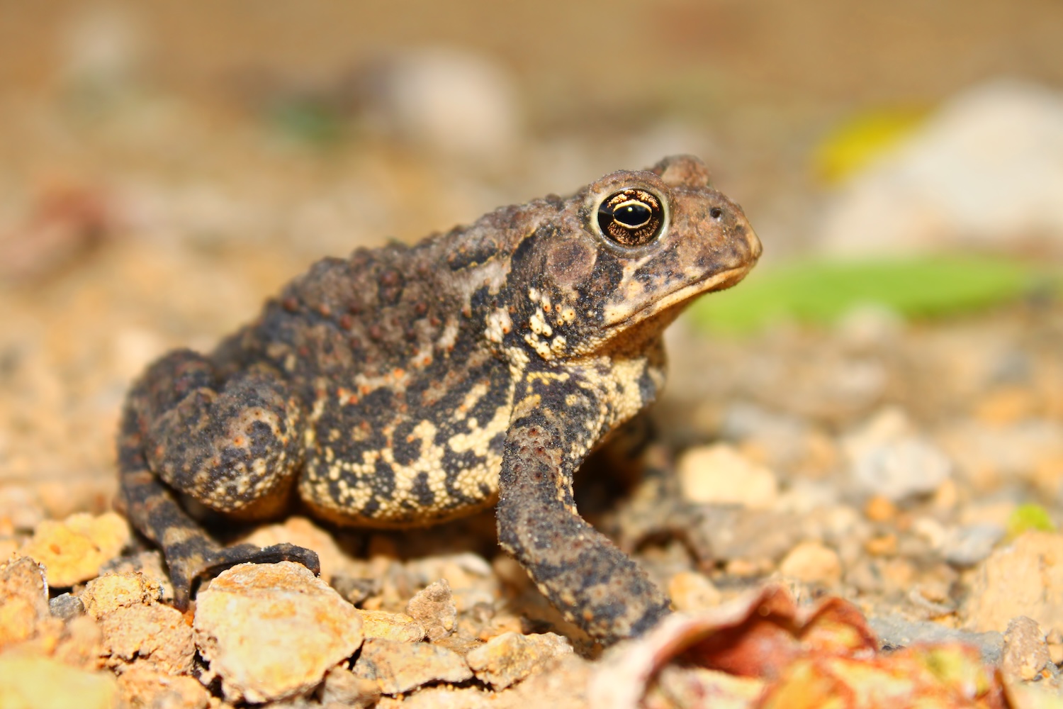
Frogs and Toads
External anatomy
Internal anatomy
Salamanders
External anatomy
Internal anatomy
Amphibian Sampling - Before You Begin
Accurate disease diagnosis depends on the collection of appropriate samples from suitable animals at the optimal stage of disease. Samples should only be collected by trained personnel using appropriate handling, restraint, and biosecurity procedures.
Suspected Reportable Pathogen
If a reportable aquatic animal pathogen is suspected, contact the appropriate state or federal aquatic animal health authority and/or the designated aquatic animal diagnostic laboratory before collecting, moving, or submitting specimens. Early communication helps ensure that appropriate sample types are collected and that regulatory, confirmatory testing, and reporting requirements are followed.
Key Biosecurity Steps
Because many amphibian pathogens can be spread through contaminated animals, equipment, water, soil, and organic debris, strict adherence to biosecurity procedures is essential.
- Wear clean disposable gloves and change gloves between animals.
- Use sterile swabs, collection tubes, and sampling equipment.
- Clean and disinfect equipment, footwear, nets, measuring devices, and handling surfaces between animal groups and sites.
- Use approved disinfectants according to manufacturer instructions, including recommended contact times.
- Avoid moving water, mud, vegetation, equipment, or animals between sites.
- Properly contain and dispose of contaminated materials
Animal Assessment and Sample Collection Tips
During health assessments and disease investigations, carefully examine the skin, eyes, oral cavity, limbs, digits, cloaca, and gills (when present). In larval amphibians, the tail and tail fin should also be evaluated. These structures are common sites for lesions and other signs of disease, including discoloration, ulceration, erosions, edema, abnormal shedding, trauma, and deformities. Body condition and hydration status should also be assessed, as changes may indicate underlying disease.
Record Situational information
For all aquatic animal disease investigations, document:
- Species and life stage
- Geographic location and date of collection
- Morbidity (illness) and mortality (death) rates, including the estimated number of animals affected.
- Clinical signs and gross lesions observed
- Environmental conditions, including water temperature, water quality parameters, habitat type, and environmental conditions.
- Relevant management history, animal movements, recent introductions, and potential exposure risks, when known.
General Sampling Tips
- Collect samples from live clinically affected animals when possible.
- Samples should also be taken from moribund animals and freshly dead specimens.
- When practical, submit whole freshly dead animals with a complete case history, as this often provides the greatest opportunity for a definitive diagnosis.
- Decomposed animals often have limited diagnostic value and should be avoided when fresher specimens are available.
- Collect samples suitable for both pathogen detection (e.g., PCR, culture, virology) and histopathology whenever possible.
- Label all specimens clearly with species, location, date, and unique animal identification.
- Store and transport samples according to laboratory recommendations and maintain appropriate temperature during shipment.
2 PHOTOS HERE
Non-Lethal Samples
- Skin Swabs: commonly used for the detection of chytrid fungi (Batrachochytrium dendrobatidis [Bd] and B. salamandrivorans [Bsal]) and other skin-associated pathogens.
- Frogs and toads: Swab the ventral abdomen, inner thighs, feet, toe webbing, and any visible skin lesions.
- Salamanders and newts: Swab the ventral surface, lateral body surface, feet, digits, tail, and any abnormal skin lesions.
- Tadpoles: Sample keratinized mouthparts (jaw sheaths and tooth rows). Additional keratinized skin may be sampled for animals nearing metamorphosis.
- If ranavirus is suspected, oral swabs and lesion swabs may also be useful.
- Toe clips and tail clips: May be collected in certain circumstances when recommended by the testing laboratory and approved by relevant aquatic animal authorities.
Necropsy Sampling
- Submit whole carcasses whenever possible, particularly for small amphibians.
- If field necropsy is necessary, collect representative samples of skin (including lesions), liver, kidney, spleen, heart, lung, gastrointestinal tract, and brain when neurologic disease is suspected.
- For histopathology, place tissues in 10% neutral buffered formalin at an appropriate tissue-to-formalin ratio.
- For pathogen detection, collect matching fresh tissues using aseptic technique and handle according to laboratory instructions.
- Whenever possible, collect duplicate samples, preserving one set in formalin for histopathology and retaining a second fresh set for ancillary testing.
When in Doubt: Contact the diagnostic laboratory before collecting or submitting samples. Laboratory personnel can provide guidance on sample selection, preservation, shipping, and appropriate testing.