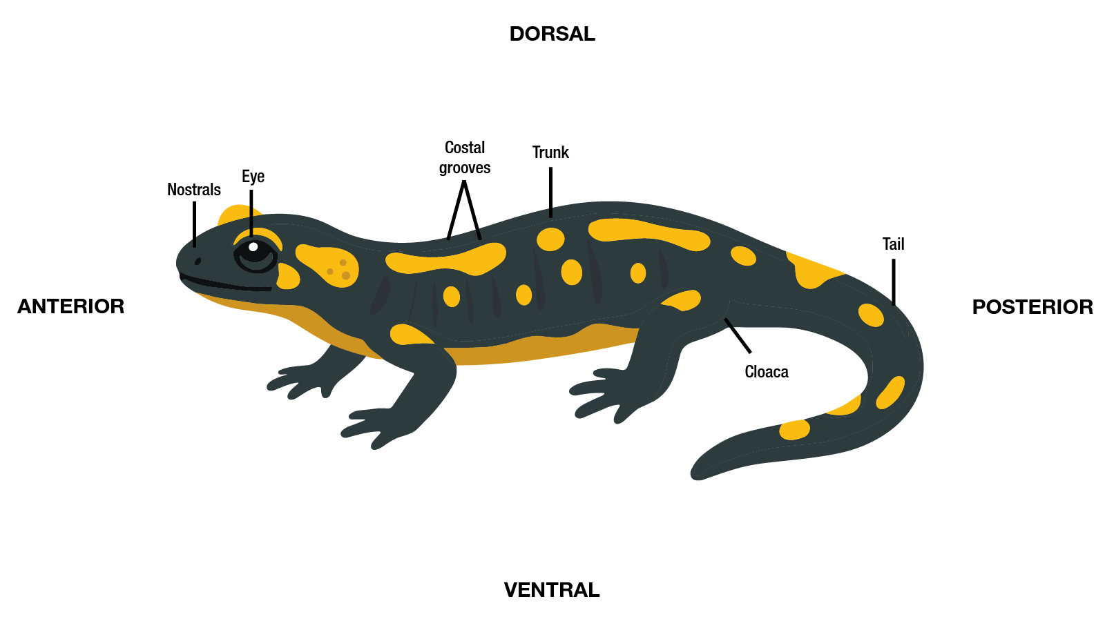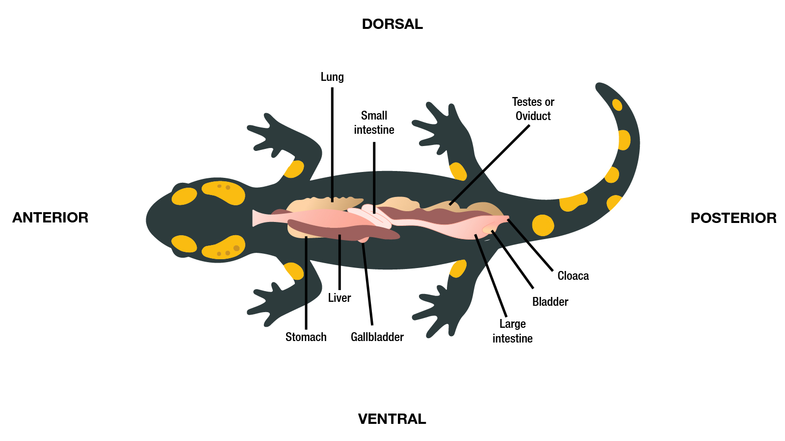
Understanding basic aquatic animal anatomy is essential for recognizing signs of disease, accurately describing lesions, and collecting appropriate diagnostic samples.
Because many pathogens target specific tissues or organ systems, knowledge of normal anatomical structures helps aquatic animal health professionals interpret lesion patterns, identify affected body systems, and conduct thorough health inspections, mortality investigations, and surveillance activities. Familiarity with anatomical terminology also promotes clear communication among field personnel, diagnosticians, producers, and regulatory officials.
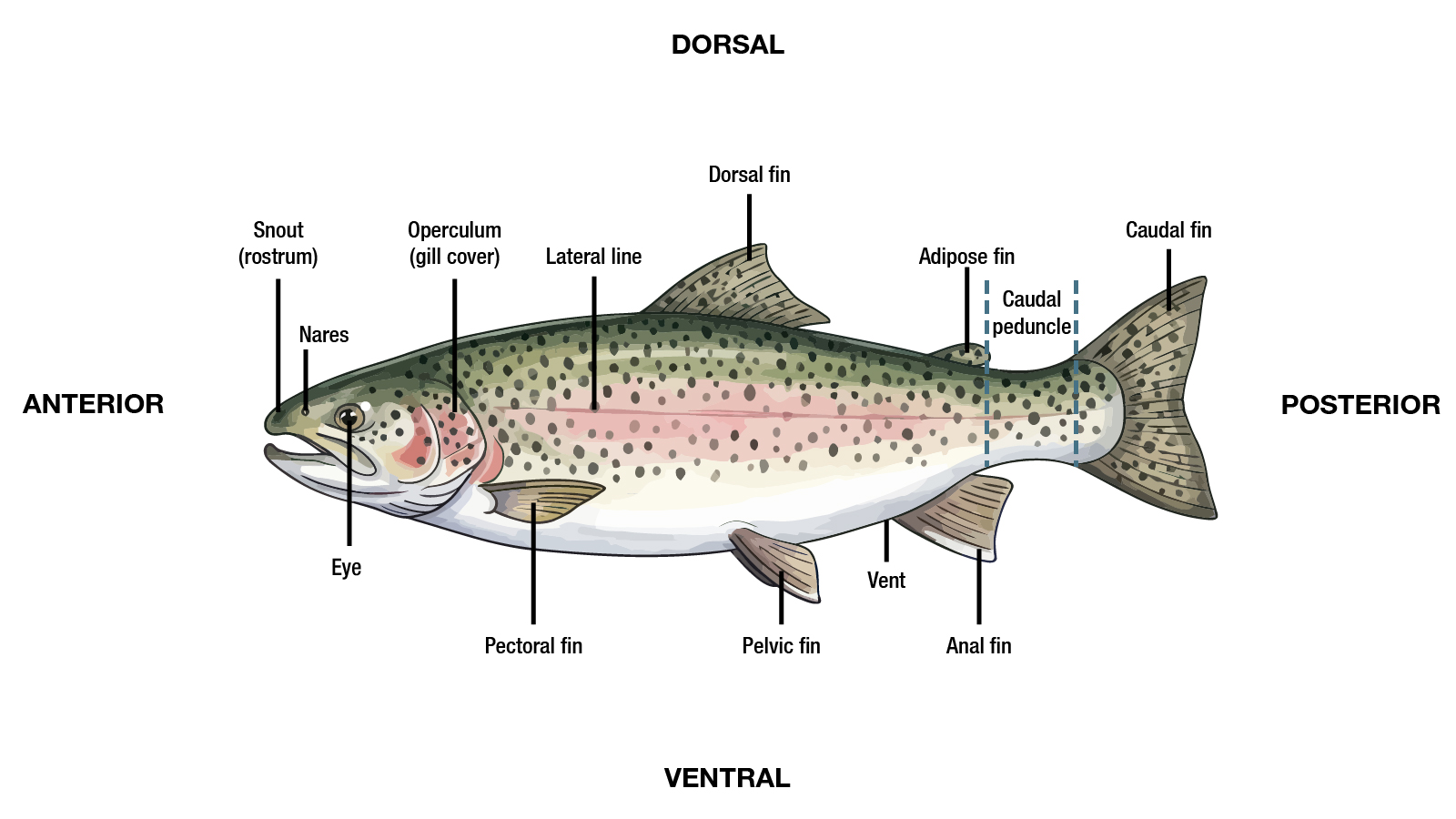
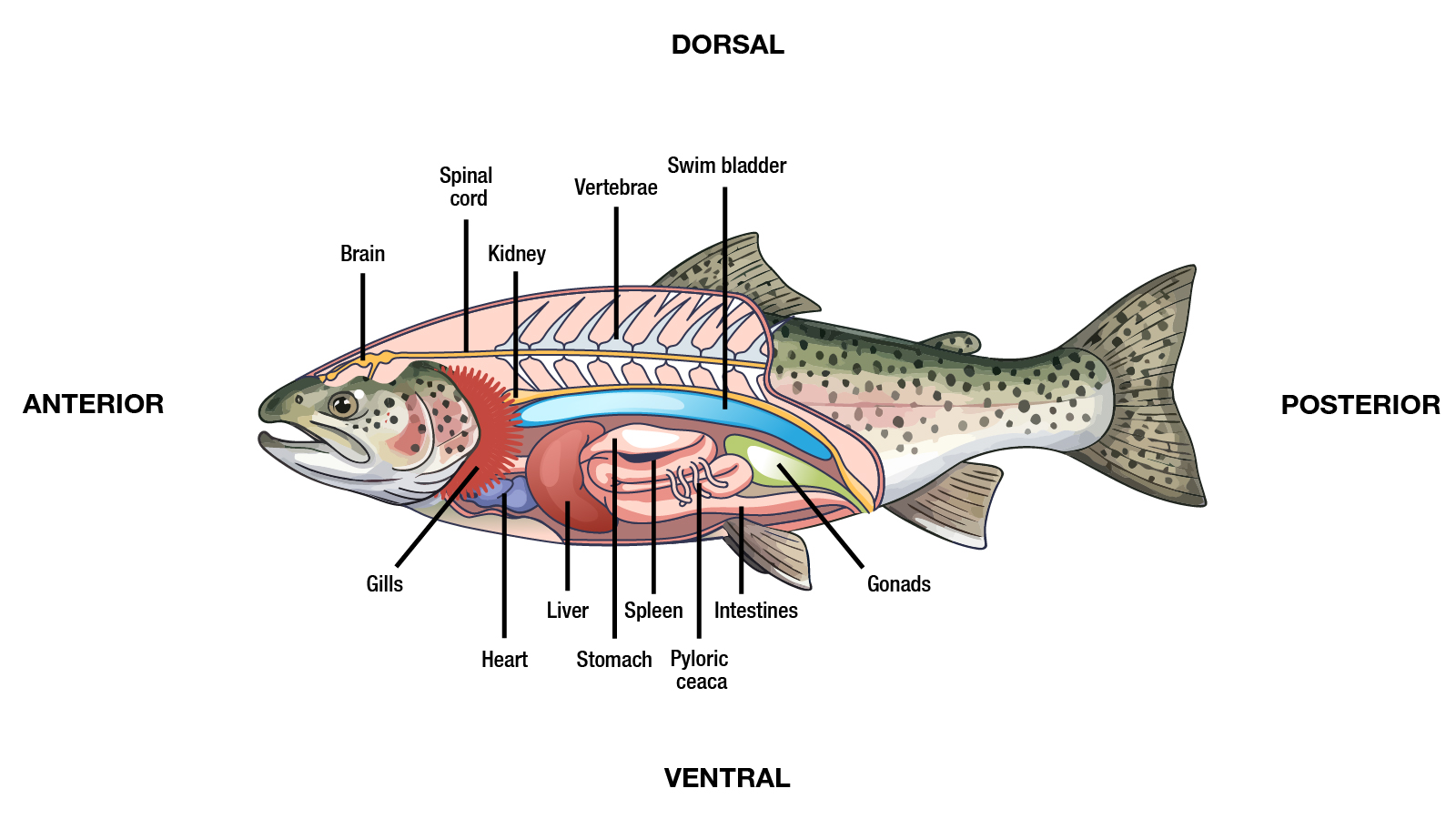
This section provides a basic overview of the anatomy of major aquatic animal groups covered in this guide, including finfish, crustaceans, mollusks, and amphibians. These illustrations are intended to support the generalized recognition of normal and abnormal findings and to aid in the localization and documentation of lesions. The focus is on structures and organs commonly evaluated in disease investigations and most frequently targeted for laboratory testing. The information is not intended to be a comprehensive anatomy reference.
Fish
The external and internal anatomy of finfish differs considerably among species.
- External variations may occur in finnage, including the number, structure (bony vs. soft), and location of fins and even eye placement as is seen in flatfishes.
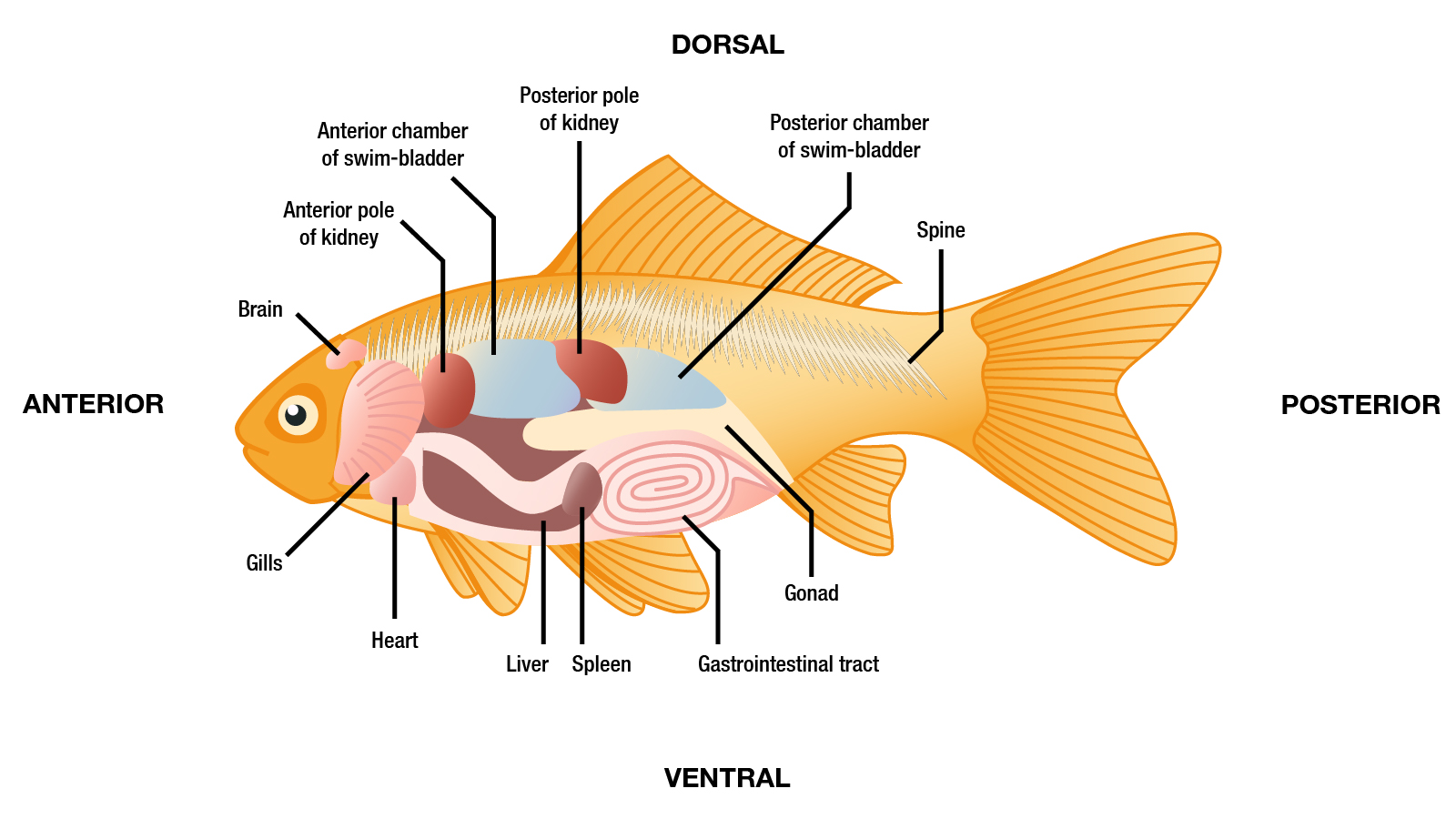
- Internal differences may include the presence or absence of a true stomach, pyloric ceca, the presence, location, and structure of the swim bladder, the length of the gastrointestinal tract (which is related to diet), and the appearance of the reproductive organs with age and stage of maturity.
- Because of these differences, it is important to understand what is considered normal for the species being evaluated.
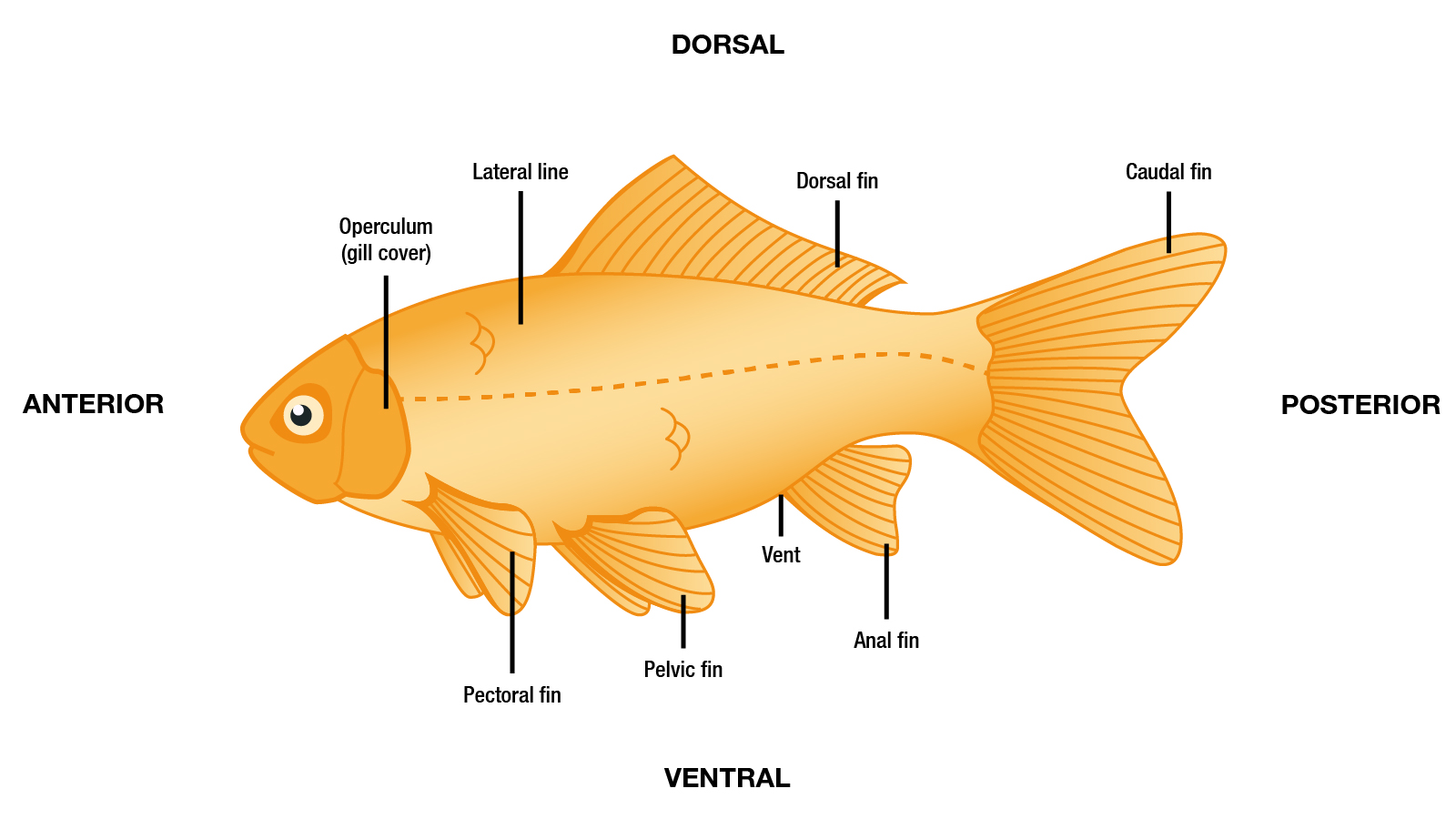
During health assessments and disease investigations, particular attention should be given to the skin, fins, gills, oral cavity, eyes, and opercula. These areas should be routinely examined, as they are common sites for visible lesions and other abnormalities.
Salmonids (salmon, trout, char) External and Internal Anatomy
Non-salmonid/Cyprinid External and Internal Anatomy
Crustaceans
The external and internal anatomy of crustaceans varies considerably among species.
- External variations may occur in body shape, exoskeleton thickness and texture, segmentation, the size and structure of appendages (such as antennae, chelae, and walking legs), and the presence and development of specialized structures used for feeding, locomotion, or reproduction.
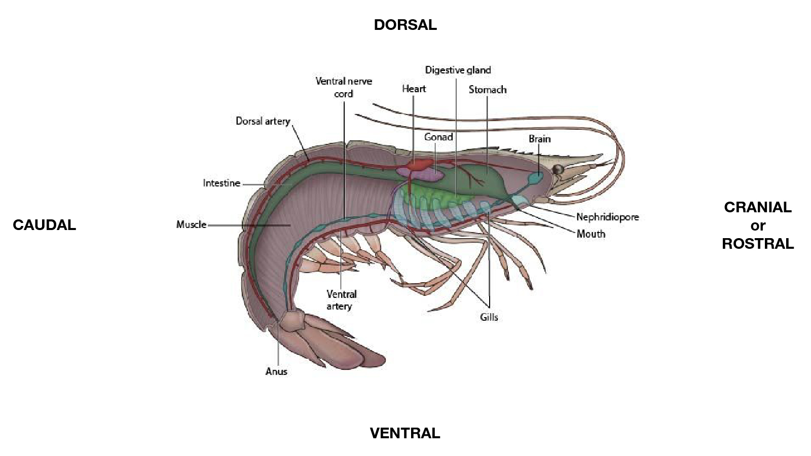
- Internal differences include the structure of the gills, the digestive system and hepatopancreas, the arrangement of the circulatory and nervous systems, and the appearance and location of reproductive organs, which vary with species, age, sex, and stage of maturity.
- Because of these differences, it is important to understand what is considered normal for the species being evaluated. Know your species before conducting a necropsy and collecting samples.
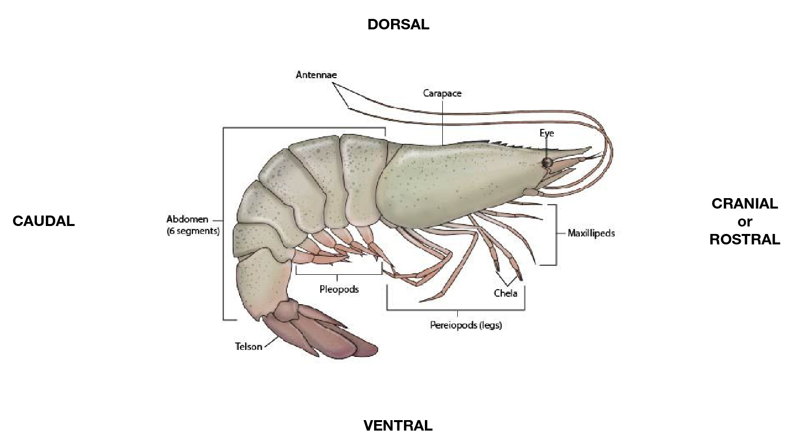
During health assessments and disease investigations, particular attention should be given to the exoskeleton (cuticle), appendages, gills, eyes, mouthparts, abdomen, and joints between body segments. These areas should be routinely examined, as they are common sites for visible lesions, discoloration, fouling, trauma, shell disease, molting abnormalities, and other signs of disease or poor health.
Shrimp External and Internal Anatomy
Mollusks
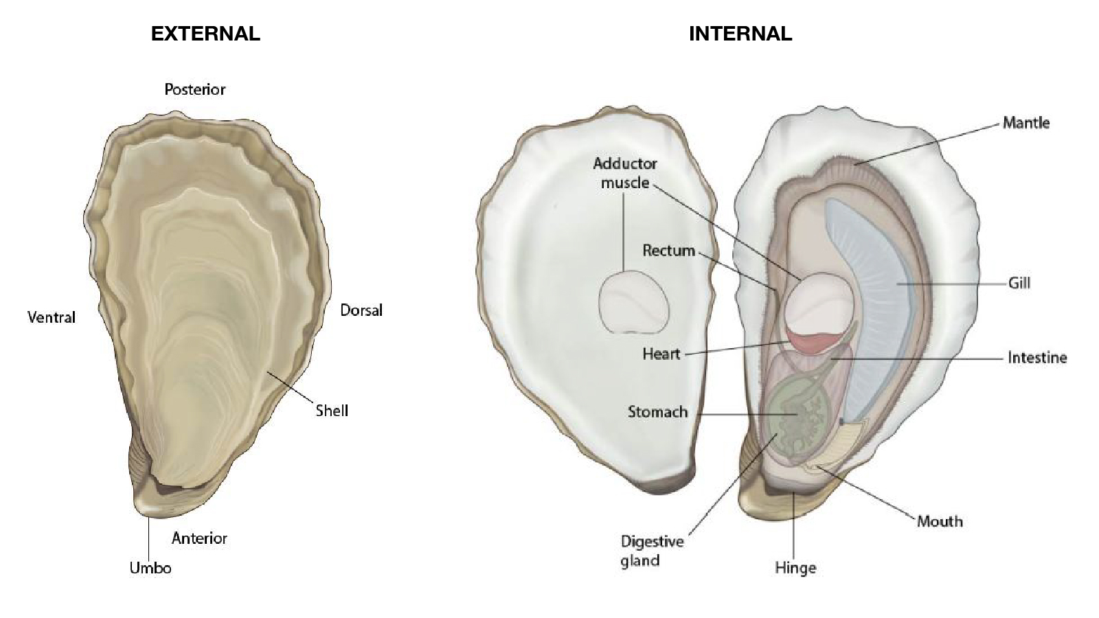
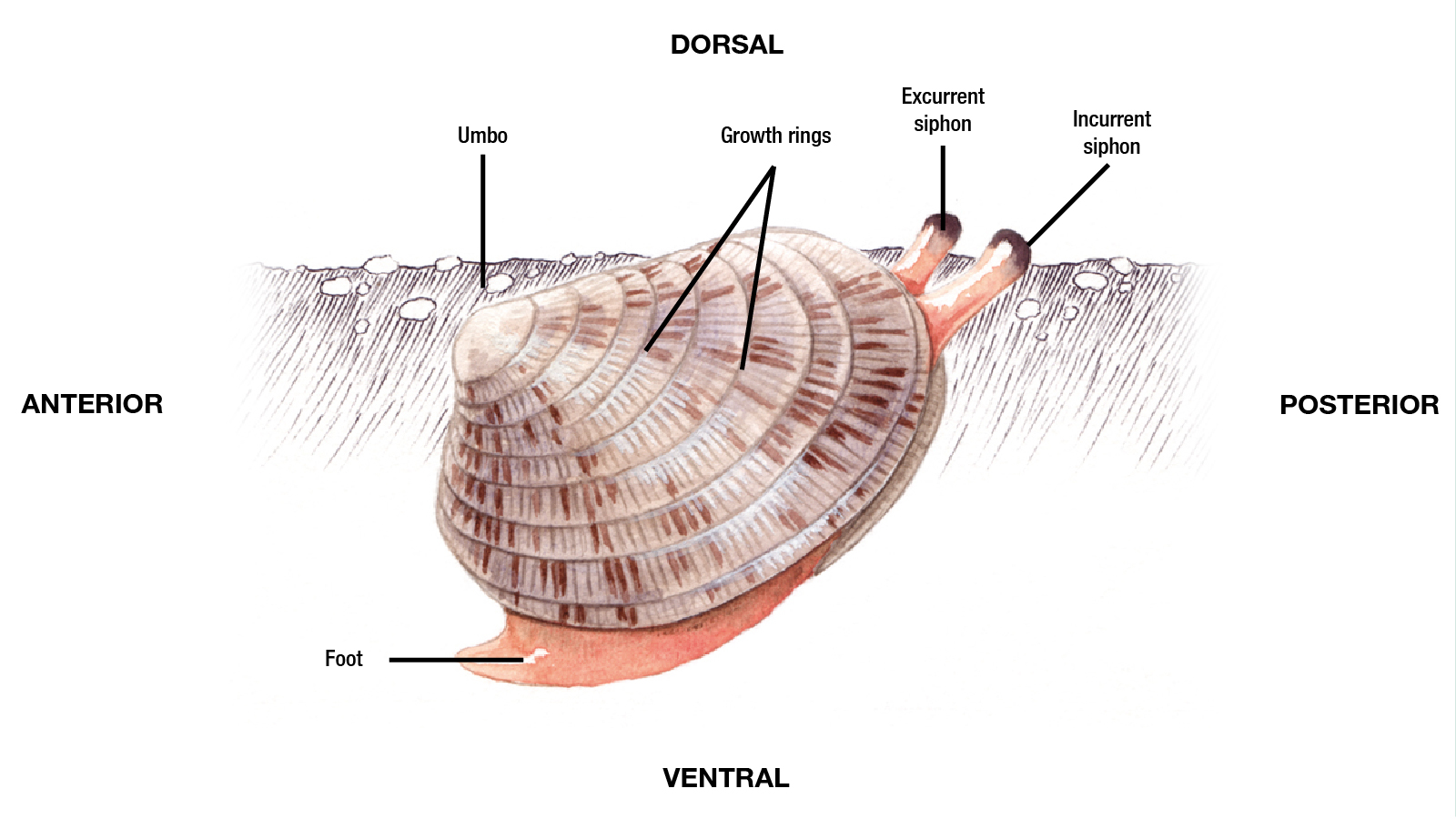
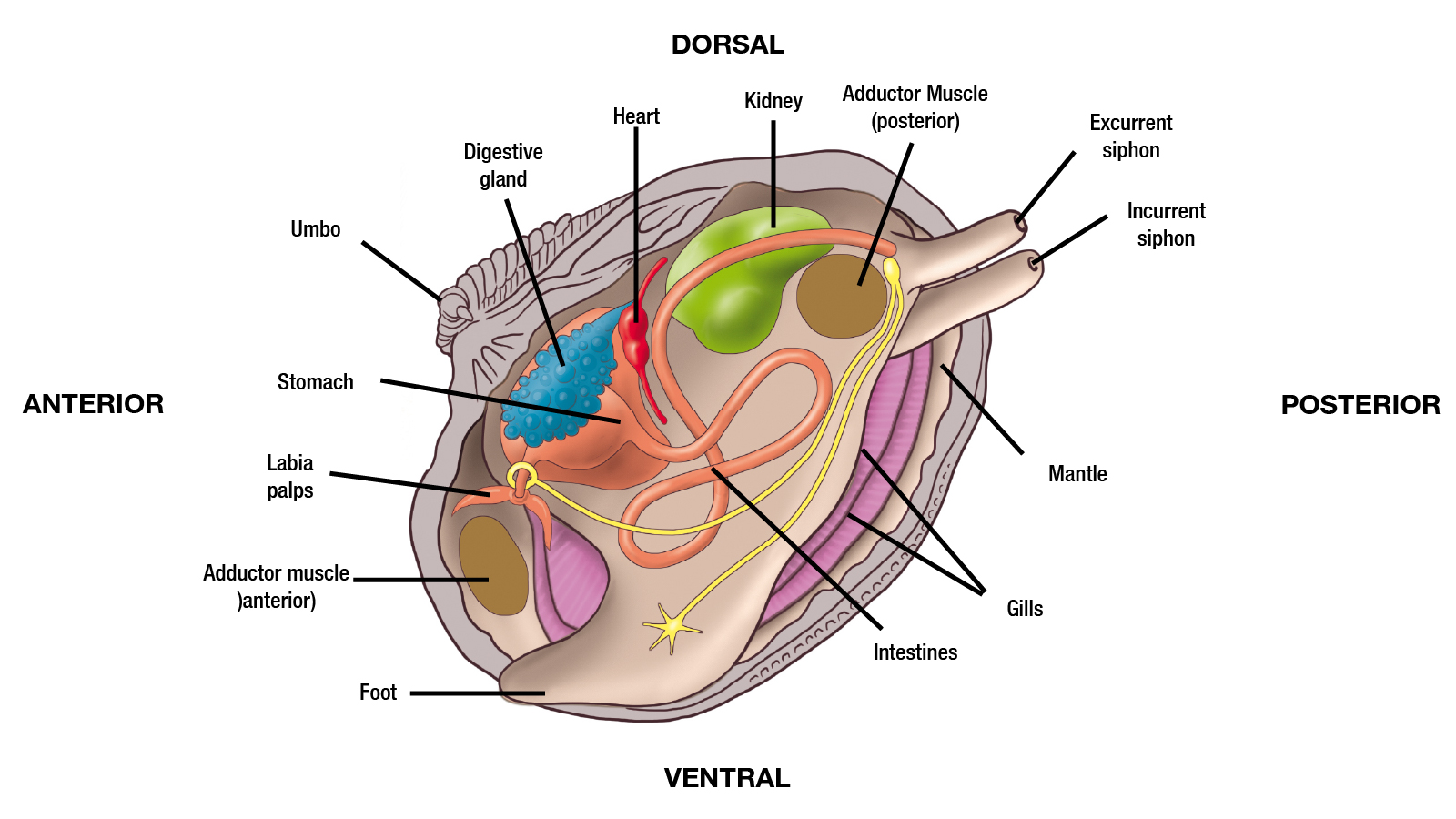
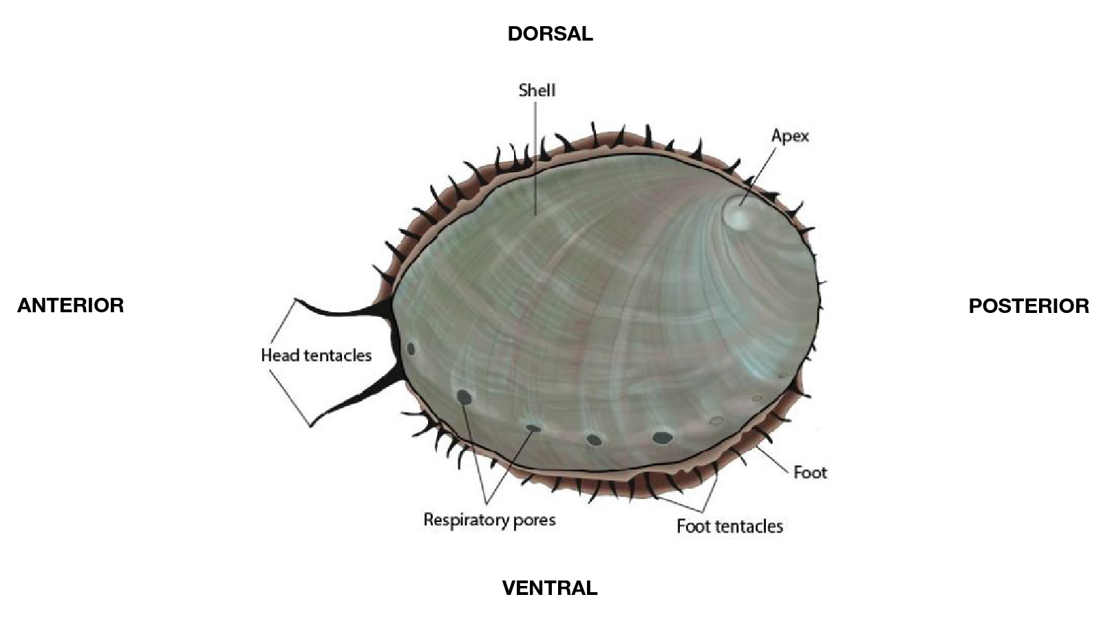
The external and internal anatomy of mollusks, including bivalves and abalone, differs considerably among species.
- External variations may occur in shell shape, size, thickness, and ornamentation, as well as in the structure of the mantle, foot, and gills. Bivalves possess two hinged shell valves and are filter feeders, while abalone have a single ear-shaped shell and a large muscular foot adapted for attachment to hard surfaces. If a bivalve shell is gaping (open) and does not close when touched, the animal is either dead or ill.
- Internal differences include the size and structure of the gills, digestive gland, gastrointestinal tract, gonads, and other organ systems. The appearance, size, and color of tissues, particularly the gonads and digestive gland, may vary with species, age, nutritional status, and stage of reproductive maturity.
- Because of these differences, it is important to understand what is considered normal for the species being evaluated. Know your species before conducting a necropsy and collecting samples.
During health assessments and disease investigations, particular attention should be given to the shell, mantle, gills, foot, digestive gland, and soft tissues. These structures should be routinely examined, as they are common sites for visible lesions, shell damage, biofouling, discoloration, tissue erosion, parasites, inflammatory changes, and other abnormalities associated with disease, environmental stress, or poor health.
Bivalve – Oyster External and Internal Anatomy
Bivalve – Clam External and Internal Anatomy
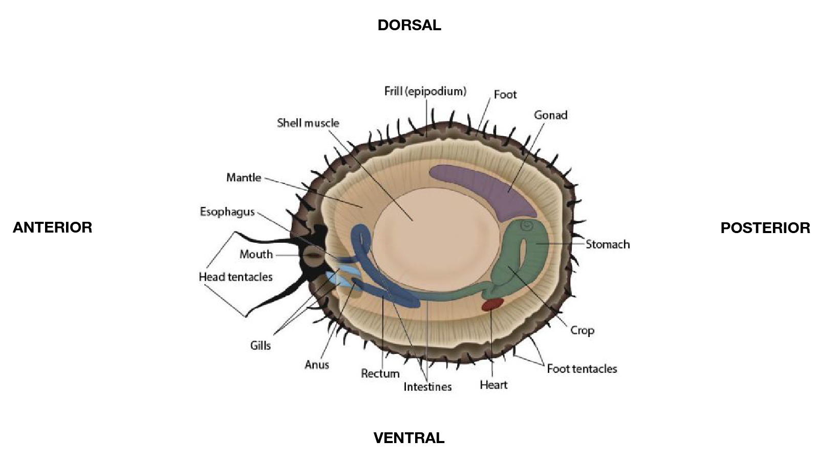
Abalone External and Internal Anatomy
Amphibians
The external and internal anatomy of amphibians differs considerably among species.
- External variations may occur in body shape, skin texture and coloration, limb size and development, and the presence or absence of structures such as external gills, tail fins, and parotoid glands.
- Internal differences include the structure of the respiratory system, digestive tract, and reproductive organs, as well as the extent of metamorphic changes that occur during development. The appearance and size of organs, particularly the gonads, may vary with species, age, sex, season, and stage of reproductive maturity.
- Because of these differences, it is important to understand what is considered normal for the species being evaluated.
During health assessments and disease investigations, particular attention should be given to the skin, eyes, oral cavity, limbs, digits, cloaca, and gills (when present). Particular care should also be taken to evaluate body condition and hydration status, as changes in these parameters may provide important indicators of underlying health problems. The tail and tail fin of larval amphibians are also important sites for detecting abnormalities. These structures should be routinely examined, as they are common sites for visible lesions, discoloration, ulceration, edema, trauma, deformities, abnormal shedding, parasitic infestations, and other signs of disease or poor health.igns of disease or poor health.
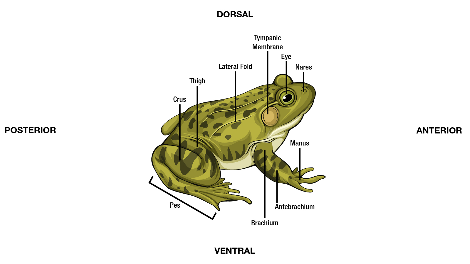
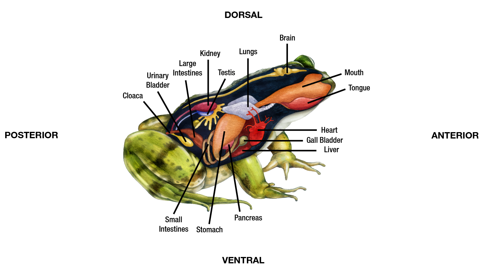
Frogs and Toads External and Internal Anatomy
Salamander External and Internal Anatomy